Please note you are entering now content intended only for Healthcare Professionals.
Are you a HCP?
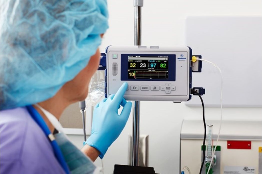
Procedural sedation often involves the use of sedative and analgesic medications like propofol and fentanyl to achieve adequate sedation and analgesia levels and ensure patient comfort. Various sedation approaches are utilized in the electrophysiology lab (EP lab). However, it's crucial to address the potential risks associated with sedation in the EP lab, such as apnea and hypoxemia.1 Patient safety is of utmost importance during procedures. To mitigate these risks, continuous monitoring of vital signs, including pulse oximetry and capnography, is essential, as adverse respiratory events are among the most common adverse events during cardiac interventions under analgosedation.2,3
Capnography, a real-time monitoring assessment of cardiopulmonary function, measures end-tidal CO2 (etCO2) levels during procedural sedation.
In today's electrophysiology lab, procedural sedation is more complex due to older patients, comorbidities, longer procedures, and deeper sedation for complex ablations. Respiratory disorders are the most common adverse events in cardiac interventions under analgosedation. A retrospective review investigated patient safety and relevant events during interventions with analgosedation in different specialties in the USA. Among adverse events (AEs) in cardiology, oversedation/apnoea (86.3%), mask ventilation (36.3%), hypoxaemia (59.1%) and administration of reversal agents due to sedative overdose (86.4%) were the most common AEs. Among all events studied, respiratory events in cardiology accounted for 68.8%.2
Capnography waveforms have proven invaluable in early detecting adverse events during procedural sedation, like airway obstruction, hypoventilation and both mild (<90% SpO2) and severe hypoxemia (<85% SpO2).4,5
Capnography detects respiratory depression at a median of 60s (5-240s) prior to pulse oximetry6 and can increase patient safety. Consequently, various international guidelines advise to monitor patients´ ventilation during procedural sedation with capnography.7 The Academy of Medical Royal Colleges (UK) recommends the use of capnography to monitor ETCO2 and ventilation in its guidelines for safe sedation with propofol during cardiac procedures8. Additionally, capnography aids in assessing the effectiveness of interventions, such as analgesia administration, to ensure optimal sedation levels are maintained9. Yet, etCO2 monitoring still appears to be underutilised in the electrophysiology lab.
In the context of electrophysiology procedures like cryoballoon ablation for atrial fibrillation (AF), capnography plays a role in monitoring cryoballoon-to-pulmonary vein (PV) occlusion. By measuring etCO2 and analyzing capnography waveforms, healthcare providers can gauge the occlusion status and evaluate pulmonary blood flow.10 Capnography monitoring during PV procedures can therefore assess the success of cryoballoon-to-pulmonary vein (PV) occlusion and monitor right phrenic nerve function. This real-time assessment has emerged as an early warning indicator11 and a predictor of long-term efficacy10 with patients that maintained normal sinus rhythm (NSR).10 Early detection of phrenic nerve injury during cryoablation procedures can help prevent potential complications early and optimize patient outcomes.
"Deep sedation with capnography monitoring is critical in achieving higher effectiveness during procedures like atrioventricular nodal re-entrant tachycardia (AVRNT) ablation, by ensuring quieter and flatter chest movements. This enhances patient comfort and minimises incidents, complications, and prolonged procedure times"
- Dr. Hammwöhner
Capnography is a valuable technology in ensuring patient safety during procedural sedation, including electrophysiology procedures like cryoablation for atrial fibrillation (AF). By incorporating capnographic monitoring, electrophysiologists can promptly detect and address respiratory complications, optimize sedation levels, and ultimately enhance patient outcomes4,5.
Don't compromise on patient safety in your electrophysiology lab during procedural sedation. Incorporate capnography monitoring, gain valuable insights in real-time, and optimize patient outcomes.
1. Tobias, J. D., & Leder, M. (2011). Procedural sedation: A review of sedative agents, monitoring, and management of complications. Saudi journal of anaesthesia, 5(4), 395–410. https://doi.org/10.4103/1658-354X.87270
2. Jones, M. R., Karamnov, S., Urman, R. D. (2018). Characteristics of Reported Adverse Events During Moderate Procedural Sedation: An Update. The Joint Commission Journal on Quality and Patient Safety, S1553725017304750. https://doi:10.1016/j.jcjq.2018.03.011
3. Bhananker, S. M., Posner, K. L., Cheney, F. W., Caplan, R. A., Lee, L. A., & Domino, K. B. (2006). Injury and liability associated with monitored anesthesia care: a closed claims analysis. Anesthesiology, 104(2), 228–234. https://doi.org/10.1097/00000542-200602000-00005
4. Gallagher J. J. (2018). Capnography Monitoring During Procedural Sedation and Analgesia. AACN advanced critical care, 29(4), 405–414. https://doi.org/10.4037/aacnacc2018684
5. Saunders, R., Struys, M. M. R. F., Pollock, R. F., Mestek, M., & Lightdale, J. R. (2017). Patient safety during procedural sedation using capnography monitoring: a systematic review and meta-analysis. BMJ open, 7(6), e013402. https://doi.org/10.1136/bmjopen-2016-013402
6. Deitch, K., Miner, J., Chudnofsky, C. R., Dominici, P., & Latta, D. (2010). Does end tidal CO2 monitoring during emergency department procedural sedation and analgesia with propofol decrease the incidence of hypoxic events? A randomized, controlled trial. Annals of emergency medicine, 55(3), 258–264. https://doi.org/10.1016/j.annemergmed.2009.07.030
7. Hinkelbein, J., Lamperti, M., Akeson, J., Santos, J., Costa, J., De Robertis, E., Longrois, D., Novak-Jankovic, V., Petrini, F., Struys, M. M. R. F., Veyckemans, F., Fuchs-Buder, T., & Fitzgerald, R. (2018). European Society of Anaesthesiology and European Board of Anaesthesiology guidelines for procedural sedation and analgesia in adults. European journal of anaesthesiology, 35(1), 6–24. https://doi.org/10.1097/EJA.0000000000000683
8. Furniss, S. S., & Sneyd, J. R. (2015). Safe sedation in modern cardiological practice. Heart (British Cardiac Society), 101(19), 1526–1530. https://doi.org/10.1136/heartjnl-2015-307656
9. Gerstein, N. S., Young, A., Schulman, P. M., Stecker, E. C., & Jessel, P. M. (2016). Sedation in the Electrophysiology Laboratory: A Multidisciplinary Review. Journal of the American Heart Association, 5(6), e003629. https://doi.org/10.1161/JAHA.116.003629
10. Pickett, R. A., Owens, K., Landis, P., Sara, R., & Lim, H. W. (2018). Cryoballoon-to-Pulmonary Vein Occlusion Assessment via Capnography Technique: Where Does Occlusion Testing by End-Tidal CO2 Measurement "Fit" as a Predictor of Long-Term Efficacy?. Journal of atrial fibrillation, 11(1), 2055. https://doi.org/10.4022/jafib.2055
11. Hoyt, R.H. & Lim, H.W. 2015. Capnographic observations during cryoballoon ablation of atrial fibrillation. The Journal of Innovations in Cardiac Rhythm Management. 6(8): 2093–2099 https://doi:10.19102/icrm.2014.060805