
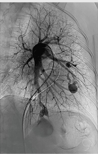
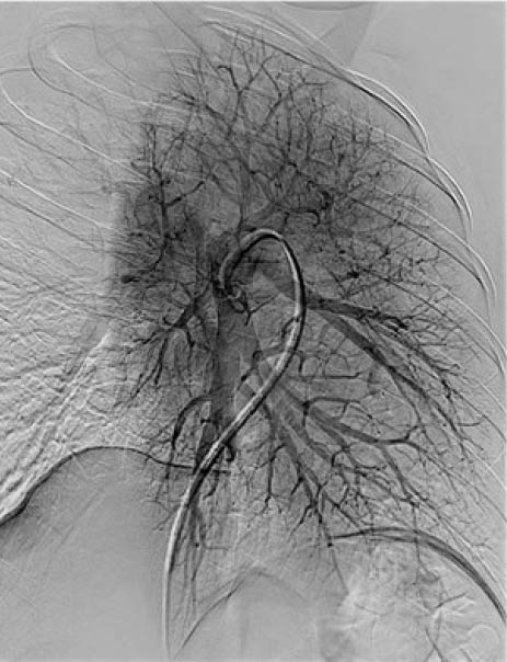
Pre angiogram
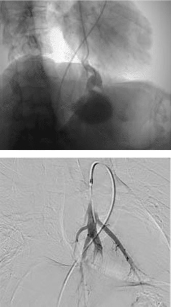
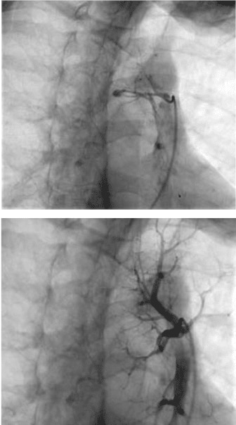
Pre and post treatment of left
lower lobe PAVM with
9mm MVP device
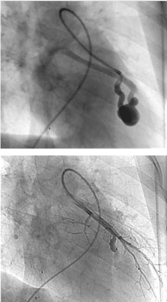
Pre and post treatment of left inferior lingular PAVM with
7mm MVP device
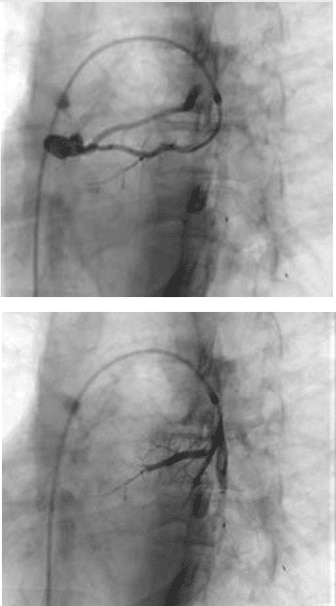
Pre and post treatment of left superior lingular PAVM with
5mm MVP device
Pre and post left lower
lobe superior PAVM with
3mm MVP device
Final angiogram
CASE DESCRIPTION
PHYSICIAN RATIONALE
VESSELS EMBOLIZED
DEVICES USED
©2023 Medtronic. All rights reserved.
⋅ 9mm MVP device
⋅ 7mm MVP device
⋅ 5mm MVP device
⋅ 3mm MVP device
Branches of the pulmonary artery
The MVP device was selected because it can be used as a single occlusion device, resulting in faster procedural times and minimal CT artifact.
The MVP devices were deployed and demonstrated immediate occlusion of each PAVM. Multiple PAVMs could be treated during the same procedure with limited radiation exposure for the patient and the operator.
34-year-old female presented for evaluation of a suspected pulmonary arteriovenous shunt, discovered on contrast-enhanced echocardiography during workup of a right posterior circulation stroke six months prior.
She reported daily epistaxis and recurrent headaches and migraines but no breathlessness or previous history of hemoptysis.
Four MVP devices were used to treat one small and three large PAVMs.
Pulmonary arteriovenous malformation (PAVM)
Pulmonary AVM


Show case

MVP™
micro vascular plug system


Back to Medtronic.com/mvp 